Unlock the potential of regenerative orthopedics in treating chronic tendinopathy for better mobility and pain relief.
Table of Contents
Introduction: Revolutionizing Chronic Tendinopathy Treatment
Welcome to Health Voice 360. I’m Dr. Alexander Jimenez, and as both a Doctor of Chiropractic (DC) and a Family Nurse Practitioner (FNP-APRN), my mission is to bridge innovative, evidence-based research and practical clinical application. In my years of practice, particularly observing patient outcomes and progressions through my clinical dashboards, I’ve seen countless individuals struggle with the debilitating pain and functional limitations of chronic tendinopathy. Conditions like tennis elbow, Achilles tendinosis, and patellar tendinopathy often become a frustrating cycle of rest, medication, and recurrence, leaving patients searching for a more definitive solution. Traditional conservative treatments, while helpful for acute inflammation, frequently fail to address the core issue in chronic cases: a degenerative, non-healing state within the tendon itself. This is where interventional and regenerative medicine offers a profound paradigm shift.
Today’s post delves into one of the most powerful and targeted techniques in my arsenal: ultrasound-guided needle fenestration. This minimally invasive procedure represents a fundamental change in our therapeutic goal. Instead of simply masking pain or reducing inflammation, we aim to strategically “wake up” the body’s own healing cascade. The core principle is to convert a chronic, stagnant, degenerative condition into an acute, controlled inflammatory event, signaling the body to initiate a robust, organized repair process. This is not about suppressing the body’s response, but about restarting it. I will guide you through the intricate physiological underpinnings of this technique, drawing on the latest findings from leading researchers pioneering its use. We will explore how modern, high-resolution ultrasound imaging has transformed this procedure from a blind “peppering” technique into a precise, targeted intervention. You will see how we can visualize diseased, hypoechoic tissue in real time, guiding a needle with sub-millimeter accuracy to disrupt the disorganized collagen fibers and calcifications that characterize tendinosis.
Throughout this comprehensive discussion, I will integrate my own clinical observations from Health Voice 360. For instance, I’ve noted that patients who undergo fenestration, especially when combined with orthobiologics like Platelet-Rich Plasma (PRP), often show faster, more sustained improvement in pain scores and functional metrics than those receiving corticosteroid injections alone. We will explore why this works—the local release of growth factors, the recruitment of fibroblasts, and the synthesis of new, healthy collagen stimulated by controlled micro-trauma. We will also discuss the synergy between fenestration and complementary injectable agents, such as hyperosmolar dextrose used in prolotherapy, which further amplifies the regenerative signal. This educational journey will provide a deep, narrative exploration of the entire process, from patient selection and pre-procedure preparation to the step-by-step execution of the technique itself, including needle positioning, in-plane and out-of-plane approaches, and how to determine the optimal number of passes. By the end, you will have a comprehensive understanding of why needle fenestration is not just a procedure, but a biologically elegant strategy to reset the healing potential of chronically injured tendons, offering new hope for lasting relief and restored function.
Understanding Tendinopathy: Beyond Simple Inflammation
As a clinician dedicated to musculoskeletal health, one of the most common and persistent conditions I encounter is tendinopathy. For decades, the suffix “-itis,” as in “tendinitis,” led both patients and practitioners to believe that the primary problem was inflammation. Consequently, the primary treatments were anti-inflammatory in nature: ice, non-steroidal anti-inflammatory drugs (NSAIDs), and corticosteroid injections. While these approaches can provide temporary relief, particularly in the very early, acute stages, they often fail to resolve the underlying issue in chronic cases. Modern histopathological research has revealed a crucial distinction.
What we are often dealing with in chronic tendon pain is not tendinitis (an active inflammatory process) but tendinosis, a degenerative condition. When we examine this tissue under a microscope, or more relevant to my clinical practice, with high-resolution diagnostic ultrasound, we don’t see the classic cellular markers of acute inflammation. Instead, we see cellular disarray and failed healing. The characteristics of tendinosis include:
- Collagen Fiber Disorganization: Healthy tendon tissue is composed of tightly packed, parallel-aligned Type I collagen fibers, giving it immense tensile strength. In tendinosis, these fibers become haphazard, thinned, and fragmented.
- Angiofibroblastic Hyperplasia: This term describes a disorganized proliferation of abnormal blood vessels (angio-) and immature tendon cells called fibroblasts (fibroblastic). These new blood vessels are often leaky and accompanied by a network of nerves, which researchers believe contributes significantly to the persistent pain of tendinopathy.
- Increased Ground Substance: The matrix between the collagen fibers becomes filled with a gel-like substance, primarily composed of glycosaminoglycans and proteoglycans. On ultrasound, this increased fluid content creates the characteristic dark, hypoechoic appearance of a diseased tendon, indicating it is less dense and structurally compromised.
- Absence of Inflammatory Cells: Crucially, there is a distinct lack of neutrophils, lymphocytes, and macrophages—the cellular hallmarks of an active inflammatory response.
This understanding is a game-changer. It explains why anti-inflammatory treatments often provide only short-term relief. They may quiet the pain-sensitive nerve endings, but they do nothing to address the fundamental structural problem of a weakened, degenerative tendon. In my clinical observations, I have seen this play out time and again. A patient with chronic tennis elbow might get a cortisone shot and feel fantastic for a few weeks. Still, the pain almost invariably returns because the underlying “sick” tendon tissue was never repaired. In fact, repeated corticosteroid injections can be catabolic, meaning they can further weaken the collagen structure and potentially increase the risk of tendon rupture down the line.
The therapeutic goal, therefore, must shift from suppressing inflammation to stimulating regeneration. We need to clear out the degenerative tissue and signal the body to lay down new, healthy, organized collagen. This is the precise rationale behind needle fenestration.
Needle Fenestration: A Regenerative Approach to Chronic Tendon Injury
Needle fenestration, also known as percutaneous needle tenotomy, is a minimally invasive procedure that embodies this new therapeutic paradigm. The term “fenestration” comes from the Latin word fenestra, meaning “window.” In this context, it refers to creating multiple small openings, or “windows,” within the substance of the diseased tendon.
The procedure is elegant in its simplicity, yet profound in its biological effect. Using a standard hypodermic needle, I repeatedly pass it through the area of tendinosis under direct, real-time ultrasound guidance. It’s crucial to distinguish this from a simple injection. While fenestration can be—and often is—combined with injecting a regenerative substance, the mechanical action of the needling itself is the primary therapeutic event.
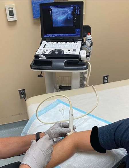
The core theory is that by causing controlled, localized micro-trauma, we are essentially converting a chronic, stagnant, degenerative condition into an acute healing response. Here’s a breakdown of the physiological cascade we aim to initiate:
- Disruption of Degenerative Tissue: The needle physically breaks up the disorganized collagen fibers and areas of calcification that characterize tendinosis. In my experience, as I perform the procedure, I can often feel the tissue texture change. The needle initially meets resistance as it enters the dense, fibrotic area. As I make multiple passes, the tissue begins to feel softer, indicating that the abnormal matrix is being broken down. This tactile feedback helps me gauge the thoroughness of the treatment.
- Initiation of a Controlled Bleeding and Inflammatory Response: Each pass of the needle ruptures the tiny, abnormal blood vessels (the “angio” part of angiofibroblastic hyperplasia). This causes localized bleeding within the tendon. While bleeding might sound counterintuitive, it is the body’s universal first step in any healing process. The resulting blood clot serves as a provisional scaffold and, more importantly, is a rich reservoir of platelets.
- Release of Growth Factors: When platelets are activated at a site of injury, they degranulate and release a powerful cocktail of growth factors. These are signaling proteins that orchestrate the entire repair process. Key players include:
-
- Platelet-Derived Growth Factor (PDGF): Stimulates cell replication and angiogenesis (the formation of new, healthy blood vessels).
- Transforming Growth Factor-Beta (TGF-?): A crucial signaling molecule that promotes the synthesis of extracellular matrix, including new collagen.
- Vascular Endothelial Growth Factor (VEGF): Promotes the formation of a new, functional blood supply to nourish the healing tissue.
- Fibroblast Growth Factor (FGF): Attracts fibroblasts to the site of injury and stimulates their proliferation.
- Recruitment of Healing Cells: These growth factors act as a clarion call, recruiting essential cells to the area. Fibroblasts, the primary cells responsible for synthesizing collagen, migrate to the site and begin the process of laying down a new matrix. This is the regenerative phase, where the body starts to build a new, stronger tendon structure.
In essence, the goal of tendon fenestration is to transform an “old,” non-healing lesion into a “hot,” active repair site. We are telling the body, “Hey, there’s an injury here that you’ve forgotten about. It’s time to fix it.” This is a fundamentally different approach from simply masking symptoms. We intervene at the cellular and molecular level to restart the body’s powerful, innate healing mechanisms.
The Crucial Role of Ultrasound Guidance in Modern Fenestration
While the concept of needling tendons has been around for some time, the advent of high-resolution musculoskeletal ultrasound has revolutionized the procedure’s safety, precision, and efficacy. Performing this technique “blind” is now considered outdated and substandard. Ultrasound guidance is not just an adjunct; it is the cornerstone of modern fenestration.
Here’s why it is essential:
- Precise Diagnosis and Targeting: Before a needle ever touches the skin, ultrasound allows me to identify and map the extent of the tendinopathy precisely. I can see the exact location of the hypoechoic (darker) and thickened areas, distinguish them from healthy, hyperechoic (brighter), fibrillar tissue, and identify any calcifications or tears. This allows me to plan my approach and ensure I am targeting only the diseased tissue while sparing the healthy portions of the tendon. For example, in the case of a tennis elbow, the pathology is often concentrated in the deep undersurface of the extensor carpi radialis brevis (ECRB) tendon at its origin. Ultrasound lets me see this with crystal clarity.
- Real-Time Needle Visualization: The most critical advantage is seeing the needle tip in real time throughout the entire procedure. Using an in-plane approach, where the ultrasound transducer is aligned with the needle’s long axis, I can visualize the needle’s entire shaft and tip as it advances through the tissue. This ensures several key safety and efficacy factors:
-
- Accuracy: I can guide the needle directly into the heart of the tendinosis, ensuring the therapeutic action is delivered exactly where it’s needed.
- Safety: I can meticulously avoid critical adjacent structures. Around the elbow, for example, are the radial and median nerves and major blood vessels. Ultrasound allows me to maintain a safe distance from these structures at all times, dramatically reducing the risk of complications like nerve injury or significant bleeding.
- Thoroughness: I can systematically work through the entire volume of diseased tissue. After making several passes in one plane, I can rotate the transducer 90 degrees into a short-axis view. This allows me to assess the medial-to-lateral extent of my treatment and redirect the needle as needed to ensure complete coverage of the treatment area.
- Dynamic Assessment: Ultrasound is not a static picture. It allows me to perform a dynamic assessment. I can have the patient gently move their wrist or ankle while I scan to see how the tendon glides and where impingement might be occurring. During the procedure, I can see the tissue’s response to the needling. I can observe disruption of the fibrotic tissue and, if I’m also injecting a fluid, see its distribution within the tendon, confirming it has been delivered to the target area.
For example, the accompanying video shows the procedure on a patient with lateral epicondylopathy, or tennis elbow. The ultrasound screen clearly shows the needle, identifiable by its bright, hyperechoic line and the characteristic reverberation artifact beneath it, advancing directly into the dark, hypoechoic area of tendinopathy within the extensor tendon. Every movement is deliberate, controlled, and guided by the live image on the screen. This level of precision is simply unattainable without ultrasound.
The Fenestration Technique: A Step-by-Step Clinical Walkthrough
Having established the “why,” let’s now delve into the “how.” A successful fenestration procedure is methodical and begins long before the treatment needle is introduced. Here is a detailed breakdown of the technique I employ in my practice, reflecting best practices from current research and my own clinical experience.
Step 1: Pre-Procedure Preparation and Anesthesia
Patient comfort and safety are paramount. The fenestration process involves multiple needle passes, which would be intolerably painful without proper local anesthesia.
- Sterile Field: First, I meticulously prep the entire area. I use a chlorhexidine or Betadine solution to create a sterile field, as in a minor surgical procedure. I cover the ultrasound transducer with a sterile probe cover and use sterile gel.
- Local Anesthesia: My preferred method is to perform a superficial skin wheal and a deeper nerve block or field block using a small-gauge needle (typically a 27-gauge) and a local anesthetic like 1% lidocaine without epinephrine. The key here is to anesthetize the skin and the subcutaneous tissues above the tendon, as well as the tendon sheath (paratenon), which is highly innervated. It is critically important not to inject the anesthetic directly into the substance of the tendon that is to be treated. The anesthetic can be toxic to tenocytes (tendon cells) and could dilute the very inflammatory and regenerative response we are trying to trigger. By placing the anesthetic around the tendon, we effectively numb the area without compromising the biological goal of the procedure.
- Vapocoolant Spray: Immediately before the main needle insertion, I often use a vapocoolant spray on the skin. This provides a brief but intense cooling sensation that acts as a counter-irritant, effectively distracting the nerve endings and making the initial needle puncture virtually painless. You can see me calling for this in the video demonstration; it’s a small detail that significantly improves the patient experience.
Step 2: Needle Positioning and In-Plane Guidance
Once the area is numb, the fenestration itself begins. I typically use a 22-gauge or 25-gauge hypodermic needle for this purpose. A slightly larger needle is more effective at disrupting tissue and is also more visible under ultrasound.
- Long-Axis, In-Plane Approach: The most common and intuitive approach is to align the needle and the ultrasound transducer along the tendon’s long axis. This is the in-plane approach: I insert the needle at a shallow angle at one end of the transducer and advance it slowly, always keeping the entire needle shaft and tip in view on the ultrasound screen.
- Setting the Trajectory: A critical pearl of the technique is to establish the correct angle of approach while the needle tip is still in the superficial subcutaneous tissue, before it ever enters the tendon. This allows me to adjust the trajectory without repeatedly piercing the sensitive tendon itself. Once I have the perfect line of sight on the ultrasound screen, aimed directly at the heart of the tendinopathy, I then advance the needle through the paratenon and into the diseased tendon tissue.
Step 3: Performing the Fenestration (The “Peppering” Technique)
This is the core therapeutic action of the procedure. The movement is not a single injection but a series of precise, repetitive motions.
- Advancing and Withdrawing: I advance the needle through the area of tendinosis, then partially withdraw it almost to the surface of the tendon, but not completely out.
- Redirecting: Before the next advance, I slightly change the needle angle, redirecting it to be either slightly shallower or slightly deeper, or fanning it out side-to-side.
- Systematic Coverage: I repeat the process of advancing, withdrawing, and redirecting sequentially. The goal is to “pepper” the entire three-dimensional volume of the tendinopathic tissue within the plane of my ultrasound view. I am methodically breaking up the degenerative matrix from multiple angles.
Step 4: Determining the Endpoint – How Many Passes Are Enough?
One of the most common questions is, “How many times do you pass the needle?” The literature provides a wide range, often suggesting between 15 and 50 passes. However, there is no magic number; it depends on the slesion’s ize and character In my practice, I rely on two key pieces of feedback:
- Tactile Feedback: As mentioned earlier, I can feel the tissue consistency changing through the needle. A fibrotic, degenerative tendon often feels gritty or tough. As I fenestrate it, it begins to feel softer and yields more easily to the needle. When the entire treated area feels uniformly soft, it strongly indicates that I have sufficiently broken up the abnormal tissue. This tactile feedback is an art that develops with experience.
- Visual Feedback: On the ultrasound screen, I am looking for subtle changes. Sometimes, I can see small areas of fluid or blood (which will appear anechoic or black) beginning to form within the tissue, signaling the start of the desired hematoma and inflammatory response. I continue the process until I am confident, based on my initial ultrasound mapping, that I have covered the entire diseased zone.
Step 5: Incorporating a Short-Axis View
After thoroughly treating the tendon in the long-axis plane, I rotate the transducer 90 degrees to obtain a short-axis or transverse view. This shows a cross-section of the tendon. From this perspective, I can confirm the medial-to-lateral extent of my treatment. If I see that I have missed a portion of the tendinopathy on one side, I can use this view to redirect the needle and ensure that the entire circumference of the lesion has been addressed.
Enhancing Fenestration: The Synergy with Prolotherapy and Orthobiologics
While needle fenestration is a powerful standalone treatment, its effects can be significantly amplified by combining it with the injection of a regenerative substance. This is a common practice, and my preferred adjunct for many cases is prolotherapy.
Prolotherapy, short for “proliferative therapy,” involves injecting an irritant solution into a ligament or tendon insertion to trigger a mild inflammatory and healing response. The most commonly used and extensively studied agent is hyperosmolar dextrose (a form of sugar).
In the video demonstration, after completing the fenestration portion of the procedure, I inject a 25% dextrose solution. Here is the rationale for this combination therapy:
- Osmotic Shock and Cell Signaling: A high concentration of dextrose creates a hyperosmolar environment outside the local cells. This causes water to rush out of the cells, resulting in cellular dehydration and stress. This stress is a powerful signal that, much like the mechanical trauma of the needling, triggers the release of local growth factors from fibroblasts and other cells. It essentially provides a secondary, chemical stimulus for the healing cascade.
- Enhanced Inflammatory Response: The dextrose solution itself acts as a mild irritant, further augmenting the inflammatory response initiated by the needling. It enhances the signal that tells the body to bring in macrophages to clean up debris and fibroblasts to start rebuilding.
- Scaffolding and Nutrient Supply: The injected fluid can also help separate tissue planes, and the dextrose can serve as a local energy source for the newly recruited cells working hard to repair the tissue.
Therefore, the combination of fenestration + prolotherapy provides a one-two punch: the mechanical disruption from the needle followed by the chemical and osmotic stimulus from the dextrose. In my clinical experience, documented through patient-reported outcome measures on our Health Voice 360 platform, this combination often leads to more robust and faster healing than fenestration alone.
Another powerful combination is fenestration with Platelet-Rich Plasma (PRP). In this procedure, we draw the patient’s own blood, centrifuge it to concentrate the platelets into a small volume of plasma, and then inject this platelet-rich solution into the fenestrated tendon. This directly delivers a supraphysiological concentration of the very growth factors that the fenestration is designed to stimulate. Fenestration with PRP is often reserved for larger tears or more stubborn cases of tendinopathy that have failed other treatments, as it represents one of the most potent regenerative strategies currently available.
Understanding Plantar Fasciitis- Video
A Clinical Case Example: Tennis Elbow (Lateral Epicondylopathy)
Let’s return to the specific case shown in the video: tennis elbow, or more accurately, lateral epicondylopathy. This condition is a classic example of tendinosis affecting the common extensor tendon at the lateral epicondyle of the humerus, particularly the extensor carpi radialis brevis (ECRB) tendon.
In the video, you see the entire procedure in action:
- The area is prepped and anesthetized.
- I use the VapoCoolant spray for patient comfort.
- I introduce the needle using an in-plane, long-axis approach, which you can see me lining up with the ultrasound transducer.
- On the ultrasound monitor, the needle appears as a bright white line advancing into the dark, thickened, hypoechoic area of tendinopathy within the ECRB tendon.
- You then see the repetitive motion of me advancing and withdrawing the needle, fanning it out to “pepper” the entire diseased region. I am physically breaking up the angiofibroblastic degenerative tissue.
- Finally, after the fenestration is complete, I inject the dextrose prolotherapy solution directly into the treated area to augment the healing response.
The entire procedure is quick, performed in the office setting, and allows the patient to go home the same day.
Post-Procedure Care and The Healing Trajectory
What happens after the procedure is just as important as the procedure itself. The healing process we have initiated takes time. It is not a quick fix.
- The Inflammatory Phase (First Few Days): Immediately following the procedure, patients can expect the area to be sore. This is a good sign; it is the desired inflammatory response kicking in. I instruct patients to avoid all anti-inflammatory medications (NSAIDs) like ibuprofen or naproxen for at least one to two weeks. Taking these would directly counteract the very biological process we worked to create. For pain management, acetaminophen (Tylenol) and ice (used sparingly for short durations) are acceptable.
- The Proliferative Phase (First 6 Weeks): This is where the magic happens. During this phase, fibroblasts actively lay down new Type III collagen, a weaker, more disorganized type of collagen that acts as a scaffold. Gentle range of motion is encouraged to prevent stiffness, but the tendon is still weak and must be protected from significant load. I typically prescribe a structured physical therapy program that begins with gentle isometrics and progresses very gradually.
- The Remodeling Phase (6 Weeks to 6+ Months): Over many months, the body will slowly remodel the weaker Type III collagen into the much stronger, more organized Type I collagen that constitutes a healthy tendon. This is where a progressive loading program guided by a physical therapist is absolutely critical. The mechanical stress of specific exercises signals the collagen fibers to align themselves properly along the lines of tension, resulting in a stronger, more resilient tendon.
I track my patients’ progress closely using validated outcome scores (e.g., Pain Visual Analog Scale, DASH for the upper extremity). What we consistently see on the Health Voice 360 dashboards is gradual but steady improvement in both pain and function over 3 to 6 months. It’s a journey, but one that addresses the root cause and offers the potential for a long-term, durable solution.
Summary
This educational post has provided an in-depth exploration of ultrasound-guided needle fenestration, a modern, evidence-based approach to treating chronic tendinopathy. We established that chronic tendon pain is typically due to tendinosis, a degenerative process, rather than tendinitis, an inflammatory one. The therapeutic goal, therefore, is not to suppress inflammation but to stimulate a regenerative healing response. Needle fenestration achieves this by using a needle to create controlled micro-trauma within the diseased tendon tissue. This mechanical disruption breaks up fibrotic, disorganized tissue and initiates a localized bleed. The resulting hematoma releases a cascade of growth factors from platelets, signaling the body to clear out debris and recruit fibroblasts to synthesize new, healthy collagen.
We emphasized the indispensable role of high-resolution ultrasound guidance, which transforms the procedure into a precise and safe intervention. Ultrasound allows for accurate targeting of the pathological tissue, real-time visualization of the needle to avoid critical neurovascular structures, and confirmation of thorough treatment. The technique itself involves an in-plane, long-axis approach, methodically “eppering” the entire volume of tendinosis until the tissue softens. The efficacy of fenestration can be further enhanced by combining it with regenerative injectables like prolotherapy (using hyperosmolar dextrose) or Platelet-Rich Plasma (PRP), which provide an additional chemical and biological stimulus for healing. Finally, we outlined the critical importance of a structured post-procedure rehabilitation program focused on protecting the tendon during the initial inflammatory and proliferative phases, followed by progressive mechanical loading during the remodeling phase to ensure the new collagen matures into a strong, functional tendon structure.
Conclusion
As a practitioner committed to leveraging the latest scientific advancements for my patients, I find needle fenestration to be one of the most elegant and effective tools for managing chronic tendinopathy. It is a paradigm shift away from the outdated model of simply managing symptoms with anti-inflammatories and cortisone shots. Instead, it is a proactive, regenerative strategy that harnesses and directs the body’s own incredible capacity for healing. By converting a chronic, degenerative lesion into an acute, controlled healing event, we are addressing the root cause of the problem at a cellular and molecular level. The combination of precise mechanical disruption under ultrasound guidance with the biological augmentation of prolotherapy or PRP offers patients who have suffered from chronic tendon pain a genuine opportunity for lasting recovery and a return to the activities they love. This approach represents the future of musculoskeletal medicine—less about suppression and more about regeneration.
Key Insights
- Tendinosis, Not Tendinitis: The foundational concept is that chronic tendon pain is a degenerative process (tendinosis) characterized by disorganized collagen and failed healing, not an active inflammatory one (tendinitis).
- Restarting the Healing Cascade: The primary goal of needle fenestration is to convert a chronic, non-healing state into an acute, controlled inflammatory response, triggering the body’s natural repair mechanisms, including the release of growth factors and the recruitment of fibroblasts.
- Ultrasound is Non-Negotiable: Modern fenestration is inseparable from high-resolution ultrasound guidance. It ensures precision in targeting diseased tissue, safety by avoiding adjacent nerves and vessels, and thoroughness in treating the entire pathological area.
- Mechanical and Chemical Synergy: The effectiveness of the mechanical disruption from the needle can be significantly amplified by the concurrent injection of a regenerative agent like dextrose (prolotherapy) or Platelet-Rich Plasma (PRP), which provides a secondary biological stimulus.
- Healing is a Process, Not an Event: Successful outcomes depend on a comprehensive approach that includes the procedure itself followed by a crucial, phased rehabilitation program. Patients must understand that healing and remodeling take several months and that their active participation in physical therapy is essential for rebuilding a strong, resilient tendon.
References
- Mishra, A. K., & Pavelko, T. (2006). Treatment of chronic elbow tendinosis with buffered platelet-rich plasma. The American Journal of Sports Medicine, 34(11), 1774-1778.
- Finnoff, J. T., twofold, S. P., & Smith, J. (2011). American Medical Society for Sports Medicine (AMSSM) position statement: interventional musculoskeletal ultrasound in sports medicine. Clinical Journal of Sport Medicine, 21(5), 369-377.
- Rabago, D., Patterson, J. J., Mundt, M., Kijowski, R., Grettie, J., Segal, N. A., & Zgierska, A. (2013). Dextrose prolotherapy for knee osteoarthritis: a randomized controlled trial. Annals of Family Medicine, 11(3), 229-237.
- Krey, D., Borchers, J., & McCamey, K. (2015). Tendon needling for treatment of tendinopathy: a systematic review. Physician and Sportsmedicine, 43(1), 80-86.
- McShane, J. M., Nazarian, L. N., & Harwood, M. I. (2006). Sonographically guided percutaneous needle tenotomy for treatment of common extensor tendinosis in the elbow. Journal of Ultrasound in Medicine, 25(10), 1281-1289.
- Jacobson, J. A. (2017). Fundamentals of Musculoskeletal Ultrasound E-Book. Elsevier Health Sciences.
Keywords
Needle Fenestration, Ultrasound-Guided Tenotomy, Tendinopathy Treatment, Chronic Tendinosis, Tennis Elbow, Lateral Epicondylopathy, Regenerative Medicine, Prolotherapy, Dextrose Injection, Platelet-Rich Plasma (PRP), Musculoskeletal Ultrasound, Collagen Remodeling, Growth Factors, Fibroblast Activation, Minimally Invasive Procedure, Alexander Jimenez, Health Voice 360
Disclaimer: This content is for educational and informational purposes only and is not intended as medical advice. The information presented here, including the techniques, procedures, and clinical observations by Dr. Alexander Jimenez, is meant to provide a general understanding of current medical concepts and should not be used to diagnose or treat any health problem or disease. It is not a substitute for professional care.
Personal Medical Disclaimer: All individuals are unique, and medical conditions present differently from person to person. You must consult with your own physician, physical therapist, or other qualified healthcare provider to obtain a proper diagnosis and to receive treatment recommendations tailored to your specific medical condition, history, and needs. Do not disregard professional medical advice or delay in seeking it because of something you have read in this post. Reliance on any information provided herein is solely at your own risk.