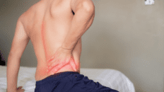
Overcome plantar fasciitis and achilles tendon issues with PRP therapy. Discover its benefits for pain and recovery.
Table of Contents
Introduction and Abstract
Welcome to our educational series. I’m Dr. Alexander Jimenez, and I hold dual credentials as a Doctor of Chiropractic (DC) and a board-certified Family Nurse Practitioner (FNP-BC, APRN). In my clinical practice, I have dedicated myself to integrating cutting-edge, evidence-based research with hands-on patient care to solve complex musculoskeletal issues. Today, we are going to explore a topic that affects millions of people worldwide: chronic heel pain, which is most commonly diagnosed as plantar fasciitis. For too long, the conventional treatment paradigm has focused almost exclusively on the plantar fascia itself—the thick band of tissue on the bottom of the foot. This often involves painful injections directly into the sole, which, as many of my patients can attest, provides temporary relief at best and can sometimes lead to further complications, such as fat pad atrophy.
In this post, I will present a paradigm-shifting approach that redirects our focus away from the symptom—the painful plantar fascia—and toward the root cause: the biomechanical dysfunction originating in the gastrocnemius-soleus complex and the Achilles tendon. Drawing upon the latest findings from leading researchers, including innovative work by orthopedic surgeons and regenerative medicine specialists, we will delve into a protocol that I have refined and successfully implemented in my own clinic. This approach is born from a deep understanding of functional anatomy and the physiological principles of tissue healing. We will discuss how chronic tightness and tendinosis (a degenerative condition, not just inflammation or tendinitis) of the Achilles tendon create a constant state of tension on the entire foot structure. This tension is particularly damaging during periods of rest, such as overnight sleep, when the foot naturally assumes a plantarflexed position, effectively shortening the calf muscles and Achilles tendon. Upon waking and taking those first steps, the tight, shortened fascia is subjected to immense tensile force, leading to the characteristic sharp, stabbing pain and the cycle of micro-tearing and chronic inflammation.

I will guide you through the intricacies of a minimally invasive, targeted injection technique designed to treat the Achilles tendon at its most vulnerable points: the musculotendinous junction (the bifurcation where the gastrocnemius muscle fibers transition into the tendon) and the enthesis (the tendon’s insertion point onto the calcaneus, or heel bone). We will explore the rationale behind using regenerative solutions like Platelet-Rich Plasma (PRP) and prolotherapy, comparing their mechanisms of action, treatment timelines, and clinical efficacy. I will share my clinical observations from years of performing these procedures, detailing the precise anatomical landmarks, needle placement techniques, and the importance of critical safety measures like aspiration to avoid intravascular injection. We will also discuss the post-procedure care protocol, including the temporary use of a walking boot to offload the tendon and a structured calf-stretching regimen to restore proper biomechanics. This post aims to provide both patients and fellow healthcare professionals with a comprehensive, modern, and evidence-based understanding of how to achieve lasting relief from chronic heel pain by treating the true source of the problem.
Rethinking Plantar Fasciitis: A Clinical Perspective
Hello, I’m Dr. Alexander Jimenez. As a clinician with credentials as both a Doctor of Chiropractic and a Family Nurse Practitioner, my practice is built on a foundation of integrated, evidence-based medicine. I constantly seek out and apply the most effective protocols to help my patients overcome debilitating conditions. One of the most common and frustrating complaints I see in my clinic is chronic heel pain, universally labeled as plantar fasciitis. For years, I, like many of my colleagues, followed the standard playbook: treat the bottom of the foot. However, through continuous research and clinical observation, it has become abundantly clear that this approach often misses the mark.
Today, I want to share a perspective that has fundamentally changed how I treat this condition—a perspective supported by emerging research from innovative orthopedic surgeons and my own extensive clinical experience. The core principle is simple yet profound: the plantar fascia is often the victim, not the culprit. The real problem frequently lies upstream, in the chronic tension and dysfunction of the gastrocnemius-soleus complex and the Achilles tendon.
In this educational overview, I will walk you through a specific, modern protocol designed to address this root cause. We will explore the anatomy, the physiological reasoning, and the practical application of this technique, moving away from painful injections in the sole to a more targeted, effective, and scientifically grounded approach.
The Anatomy of the Problem: Identifying the True Source of Heel Pain
To effectively treat chronic heel pain, we must first understand the biomechanical chain that leads to the pathology. The traditional focus on the plantar fascia—that thick, fibrous band of connective tissue running along the bottom of the foot from the heel to the toes—is a focus on the site of pain, not necessarily the source of the dysfunction.
Think of the foot and lower leg as a functional unit. The powerful gastrocnemius and soleus muscles in the calf merge to form the Achilles tendon, the strongest and thickest tendon in the human body. This tendon inserts onto the posterior aspect of the calcaneus (the heel bone). On the underside of this same bone, the plantar fascia originates. This creates a biomechanical “tug-of-war.”
When the gastrocnemius-soleus complex is chronically tight, a condition often exacerbated by modern lifestyles involving prolonged sitting or improper footwear, it exerts a constant tensile force on the Achilles tendon. This chronic tension leads to a degenerative condition known as tendinosis. It’s crucial to distinguish tendinosis from tendinitis.
- Tendinitis is an acute inflammatory condition, characterized by inflammatory cells and the classic signs of inflammation (redness, swelling, heat, pain). It typically responds well to rest, ice, and anti-inflammatory measures.
- Tendinosis, on the other hand, is a chronic, degenerative state. Histologically, we see a disorganization of collagen fibers, an increase in ground substance, neovascularization (abnormal new blood vessel formation), and a notable absence of inflammatory cells. This is a failed healing response. The tissue is weakened, dysfunctional, and painful.
This chronic Achilles tendinosis is the engine driving plantar fasciitis. When you are at rest, especially during sleep, your foot naturally falls into a position of plantarflexion (pointing downwards). This allows the already-tight calf muscles and Achilles tendon to shorten even further. The plantar fascia, in this relaxed state, also shortens.
The classic symptom of plantar fasciitis—excruciating pain with the first steps in the morning—is the direct result of this cycle. When you stand up, your body weight suddenly forces your foot into dorsiflexion, rapidly and forcefully stretching the shortened, dysfunctional, and vulnerable plantar fascia. This abrupt stretch causes micro-tears at the fascia’s insertion point on the calcaneus. Each step repeats this traumatic event, perpetuating a cycle of injury, pain, and failed healing. The pain you feel is the cumulative effect of thousands of these micro-tears. By addressing only the plantar fascia, we are merely managing the symptom while ignoring the relentless pull from the Achilles tendon that causes the injury day after day.
A Paradigm Shift in Treatment: Targeting the Achilles Tendon
Based on this understanding, the logical and most effective treatment strategy is to intervene at the source of the tension: the Achilles tendon. The goal is to release the chronic tightness and stimulate a proper healing response within the degenerative tendon tissue. This is achieved through a targeted injection protocol that addresses two critical anatomical locations.
Injection Site 1: The Musculotendinous Junction (Bifurcation)
The first and most critical injection point is the musculotendinous junction of the gastrocnemius. This is the area where the muscle fibers of the calf begin to transition into the dense, fibrous tissue of the Achilles tendon. We refer to this as the bifurcation. This zone is a common site of mechanical stress and degenerative change.
Identifying the Landmark:
To locate this point accurately, the patient should be positioned prone on the examination table with their foot hanging off the edge. This allows for easy manipulation and palpation. I find the easiest way to pinpoint the location is to relax the patient’s leg completely. I then place their foot against my leg or use my hand to dorsiflex it passively. This puts a slight tension on the tendon, making it more prominent.
I run my thumb up the midline of the Achilles tendon from the heel. As I move superiorly, I can feel the distinct, cord-like tendon. I continue this path until I feel a subtle change in the tissue texture. The firm, ropey tendon will split or “bifurcate” into the softer, more muscular bellies of the medial and lateral heads of the gastrocnemius. This bifurcation point is our target. In most individuals, it feels like a small divot or softening right at the apex where the two muscle bellies meet the tendon.
- Clinical Note: My table at the clinic is stationary, which provides a stable platform. On a rolling table, like the one we’re using for this demonstration, extra care must be taken to stabilize both the patient and myself. I ask the patient to flex their foot (dorsiflexion). “Flex that foot. There you go.” This action tenses the entire complex, making the bifurcation even more palpable. “And then run up that tendon till you find it. Oh! Right there, feel it?” The patient will often confirm the tenderness at this exact spot. This tenderness is a clinical sign of the underlying tendinosis. That’s your entry point.
The Injection Technique:
For this procedure, we use a syringe with a specific volume of regenerative solution. Let’s say we have five cubic centimeters (cc’s) of solution. We need to allocate this volume strategically across all injection sites. For this first site, we will use approximately 1- 2.5 cc, depending on the total volume available and the number of sites we plan to treat.
- Preparation: The skin is meticulously cleaned with an antiseptic solution like alcohol or chlorhexidine to minimize the risk of infection.
- Needle Entry: The needle, with the bevel pointing up, is inserted at the identified bifurcation point. The initial entry is perpendicular to the skin, just enough to break through the subcutaneous tissue.
- Repositioning and Advancement: Once through the skin, the angle of the needle is changed. I drop the syringe down so the needle is angled more parallel to the long axis of the tendon. “You’re going to break sub-Q and then drop it down.” The goal is to get into the substance of the tendon, not just around it. Remember, the tendon is a thick structure here. You have to get about half the length of a standard 1.5-inch needle in.
- Advancing into the Tendon: I then carefully advance the needle into the tendinous tissue. “Then lean it and run it into the tendon.” As the needle enters the dense, fibrotic tissue of the degenerative tendon, there is a distinct change in resistance. It feels different from passing through healthy muscle or fat. “Once you’re there, you’ll feel it change. You’ll go, ‘Okay, I’m there.'” This tactile feedback is crucial for confirming correct placement.
- Injection: Once I’m confident the needle tip is within the substance of the tendon at the musculotendinous junction, I slowly inject the solution—about one cc. The injection should be done gently to allow the solution to permeate the dense tissue. It’s important to be gentle with the patient and communicate throughout the process. “Did you feel the tissue change? No pain at all? Yes, I got you.” The patient’s feedback is invaluable.
It’s important to use the right tool for the job. In my office, I typically use a 1.5-inch needle for this procedure. The longer needle allows me to enter the skin and then lay the needle down almost parallel to the leg, ensuring the tip can be maneuvered deep within the tendon. If you use a shorter needle and try to lay it down, the tip may remain in the subcutaneous tissue, rendering the injection ineffective. You must get deep to the subcutaneous layer to reach the target.
Injection Site 2: The Achilles Tendon Insertion (Enthesis)
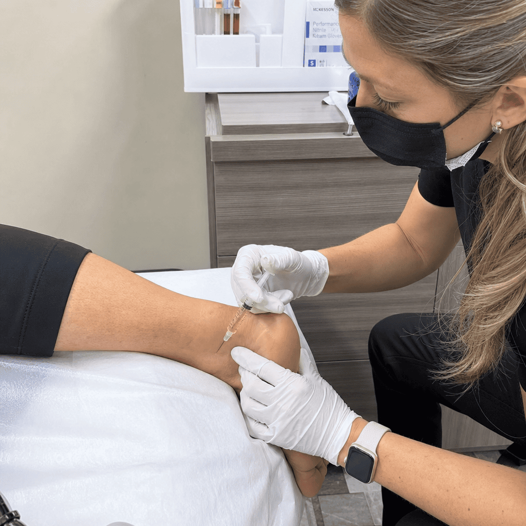
The second key area to treat is the enthesis, which is the site where the Achilles tendon inserts directly onto the calcaneus. This is another area of high mechanical stress and is frequently involved in the pathology of tendinosis, often referred to as insertional Achilles tendinopathy.
Identifying the Landmark:
With the patient still in the prone position, I palpate the posterior aspect of the heel. The insertion of the Achilles tendon is a broad “fan” of tissue that attaches to the bone. I carefully feel along the medial (inner) and lateral (outer) borders of this insertion. I ask the patient, “Is it more tender medial or lateral?” Very often, patients will report significantly more tenderness on the medial side. “Medial for sure. Medial, yeah, medial.” This is a common finding and helps guide the injection to the area of maximum pathology.
Our target is the precise point of attachment, right on the bone, where the tenderness is greatest. “So you find right where the tendon attaches… right there.”
The Injection Technique:
- Angle of Approach: For this injection, I approach from a slightly angled trajectory, coming in towards the bone.
- Depth: The goal is to reach the bone-tendon interface. I advance the needle until I feel it make gentle contact with the periosteum (the outer surface of the bone). This confirms the correct depth. We only need to go in about half the needle’s length.
- Aspiration: A Critical Safety Step: Before injecting any substance at this location, or any location for that matter, it is absolutely imperative to aspirate. This means pulling back on the plunger of the syringe for a few seconds to create negative pressure. “Now if you’re doing this with prolotherapy, please for the love of Jesus aspirate before you inject.”
-
- Why is this so important? The area around the heel is well-vascularized. If the needle tip has inadvertently entered a blood vessel, aspirating will draw blood back into the hub of the needle, creating a “flash.” “If you get a flash, you know you’re in the wrong place.”
- What happens if you inject into a vessel? The local anesthetic commonly used in these solutions (like lidocaine or procaine) can have serious systemic effects if injected intravenously. It can cause cardiac arrhythmias, seizures, or other central nervous system toxicities. “If you put lidocaine into the vessel… then you got a problem. You’re going to have a sleepover that you didn’t plan for.” It’s a medical emergency.
- What to do if you get a flash? If blood appears in the hub, do not inject. I immediately withdraw the needle, apply pressure to the site, and then clear the needle of blood by pushing the plunger slightly. I then find a new trajectory to re-insert the needle and aspirate again before proceeding.
-
- Injection: Once aspiration is negative (no blood return), I inject a small volume of the solution, typically around half a cc, directly onto the enthesis. If both the medial and lateral sides are tender, I will administer half a cc to each spot.
The Choice of Regenerative Solution: Prolotherapy vs. PRP
The solution we inject is designed to stimulate the body’s natural healing cascade. Two of the most effective options in my clinical practice are prolotherapy and Platelet-Rich Plasma (PRP).
Prolotherapy: Stimulating a Healing Response
Prolotherapy, short for “proliferative therapy,” involves injecting a solution—most commonly a dextrose (sugar) solution mixed with a local anesthetic—into degenerative or injured connective tissues like tendons and ligaments.
- Mechanism of Action: The dextrose solution acts as a mild irritant. It creates a localized, controlled inflammatory response. This is key because, as we discussed, tendinosis is a condition with a lack of inflammation. By inducing this acute inflammation, we are essentially “waking up” the body’s healing machinery. This new inflammatory cascade brings growth factors, fibroblasts, and other healing cells to the area. These cells then lay down new, healthy, and organized collagen fibers, effectively remodeling and strengthening the weakened tendon.
- Treatment Protocol and Efficacy: Prolotherapy is a highly effective and cost-efficient treatment. However, its effects are cumulative. “The thing with prolotherapy is it’s going to take about six treatments to start seeing significant turnaround.” A typical protocol in my clinic involves weekly injections for the first four weeks, followed by two more treatments every other week. By the end of this six-treatment course, patients generally show significant and lasting improvement.
One of the participants in our session shared a personal experience that highlights a common pitfall: “I was going prolotherapy on myself right into the fascia… I stopped because it just sucks… now you have this new injury from the needle in my fascia.” This is a critical point. Injecting a proliferant like dextrose directly into the already-traumatized plantar fascia can be counterproductive. You are adding a new inflammatory trauma to a sensitive tissue that is already struggling to heal. This is why our protocol focuses on the Achilles tendon—we are treating the cause, not adding to the injury at the site of the symptom.
Platelet-Rich Plasma (PRP): Concentrating the Body’s Healing Power
PRP is a more advanced form of regenerative medicine. The process involves drawing a sample of the patient’s own blood and spinning it in a centrifuge to separate and concentrate the platelets. This platelet-rich plasma, which contains a potent cocktail of natural growth factors, is then injected back into the injured area.
- Mechanism of Action: Platelets are the first responders to injury. When activated, they release a host of powerful growth factors, including Platelet-Derived Growth Factor (PDGF), Transforming Growth Factor-beta (TGF-beta), Vascular Endothelial Growth Factor (VEGF), and others. These growth factors are the master signals that orchestrate the entire healing process. They recruit stem cells, stimulate cell proliferation, promote new blood vessel formation (angiogenesis), and direct the synthesis of a new collagen matrix.
- Treatment Protocol and Efficacy: Because PRP delivers a much higher concentration of growth factors directly to the site of injury, it is generally more efficient and powerful than prolotherapy. “PRP… works just as good [as prolotherapy]. However… prolotherapy is not as efficient as PRP.” Often, a single PRP treatment, or perhaps a series of two to three treatments, can achieve results that might take six or more prolotherapy sessions. The choice between PRP and prolotherapy often comes down to the severity of the condition, patient preference, and cost considerations. For particularly stubborn cases or for patients seeking a faster recovery, PRP is my preferred method. As I often say in the clinic, “Show me something tender, and I’ll show you my favorite place to inject PRP.”
The Genesis of the Protocol: A Story of Clinical Innovation
This protocol wasn’t developed in a vacuum. It was born from clinical frustration and collaborative innovation. I want to share the story of how it came to be, as it illustrates how clinical science advances.
About eight years ago, I was discussing the persistent challenge of treating plantar fasciitis with a colleague, Dr. Roman, an orthopedic surgeon who is also on the faculty at the University of Nevada Medical School. We both felt that the standard treatments were failing too many of our patients. He expressed his immense frustration, particularly with a patient he was treating that very day. He had tried everything, including a fat pad replacement surgery, a procedure attempted when chronic steroid injections have caused fat pad atrophy in the heel. Steroids, while providing temporary anti-inflammatory relief, can degrade the natural fatty cushion of the heel over time, leading to a new and often permanent source of pain known as fat pad syndrome. “When you inject the fat pad in the heel long enough, it goes away… And that fat never comes back.” His attempts to fix this patient’s pain, even with surgical interventions, were unsuccessful.
That evening, we sat down and, fueled by a couple of Moscow Mules, we started sketching on bar napkins. I posed the question: “Roman, I got to have a better way to treat plantar fasciitis.” He immediately replied, “We got to stop sticking needles in the bottom of our people’s feet.” We mapped out the entire biomechanical chain, from the gastrocnemius down to the plantar fascia. We theorized that by releasing the tension in the Achilles tendon, we could give the plantar fascia the mechanical rest it needed to heal finally. We concluded, “There’s no reason this won’t work.”
We both started implementing this new Achilles-focused protocol in our respective practices. The results were immediate and dramatic. It was, as I’ve come to describe it, “crazy good.” Because of his academic position, Dr. Roman was able to formalize this work. He began by publishing an N-of-one study (a detailed case study of a single patient), which is often the first step in introducing a new clinical technique to the scientific community. Subsequently, he and his team at the medical school ran larger clinical trials, which validated the efficacy of the approach. While this technique is still not widely disseminated in the mainstream literature, its clinical success is undeniable. The story of its development is a testament to the power of interdisciplinary collaboration and questioning established dogma to find better solutions for our patients.
Post-Procedure Care: The Path to Lasting Recovery
The injection is only the first step. Proper post-procedure care is essential to ensure the healing process is successful and the results are long-lasting.
- The Walking Boot: Immediately following the procedure, I place the patient in a cam walker boot. The purpose of the boot is to offload the Achilles tendon and the plantar fascia. By immobilizing the ankle, the boot prevents the constant stretching and re-injury cycle, giving the newly stimulated tissues a protected environment in which to heal. I typically have patients wear the boot for three days. This short period of immobilization is critical.
- Therapeutic Stretching: After the initial three-day period in the boot, the patient begins a structured calf stretching program. This is not just casual stretching; it is a therapeutic intervention designed to lengthen the gastrocnemius-soleus complex and restore normal biomechanics permanently. The goal is to address the underlying tightness that caused the problem in the first place. These stretches must be performed consistently and correctly to prevent the recurrence of tension and pain.
By combining the targeted regenerative injections with this post-procedure protocol, we are not just treating the pain; we are correcting the underlying biomechanical fault. We are fixing the tendinosis in the Achilles tendon, which in turn allows the damaged plantar fascia to heal.
As I’ve explained to countless patients, the pain from plantar fasciitis is not a disease of the fascia itself in most chronic cases. It’s a symptom of a larger mechanical problem. We fix the Achilles, and the plantar fascia has a chance to heal. The only time the plantar fascia itself is the primary problem is typically in cases of acute trauma—for instance, stepping on a sharp object, landing hard from a jump, or forcefully overstretching and tearing the tissue. But for the vast majority of chronic sufferers, the pathway to relief lies in the calf and the Achilles tendon.
Summary, Conclusion, and Key Insights
Summary
This educational post, presented from my perspective as Dr. Alexander Jimenez (DC, FNP-APRN), outlines a modern, evidence-based protocol for treating chronic heel pain, commonly known as plantar fasciitis. The central thesis is a paradigm shift away from treating the plantar fascia directly and instead focusing on the root biomechanical cause: chronic tendinosis and tightness within the gastrocnemius-soleus-Achilles complex. Constant tension from a dysfunctional Achilles tendon places the plantar fascia under relentless strain, leading to a cycle of micro-tearing and failed healing that manifests as sharp heel pain, particularly with the first steps in the morning.
The proposed treatment involves targeted regenerative injections at two key anatomical sites: the musculotendinous junction (bifurcation) of the gastrocnemius and the Achilles tendon insertion (enthesis) on the calcaneus. We discussed the precise techniques for locating these points and performing the injections, emphasizing the critical importance of tactile feedback and safety procedures like aspiration to prevent intravascular injection. We compared the mechanisms and efficacy of two primary regenerative solutions, prolotherapy and Platelet-Rich Plasma (PRP), noting that while both are effective, PRP offers a more potent and efficient healing response. The post-procedure protocol, consisting of a short period of immobilization with a walking boot followed by a dedicated calf stretching regimen, is highlighted as an essential component for achieving long-term success by correcting the underlying mechanical dysfunction.
Conclusion
The approach detailed in this post represents a significant evolution in the management of chronic heel pain. By understanding and addressing the upstream cause—the degenerative Achilles tendinosis—we can provide patients with a more effective, logical, and lasting solution than the traditional, often painful and ineffective, method of injecting the sole. This protocol, born from clinical collaboration and supported by growing evidence and years of successful application in my practice, empowers clinicians to break the cycle of chronic injury and facilitate true healing. It underscores the principle that effective treatment requires a deep understanding of functional anatomy and a willingness to challenge conventional wisdom in pursuit of better patient outcomes. The future of musculoskeletal medicine lies in these targeted, regenerative approaches that treat the source of the pathology, not just the location of the pain.
Key Insights
- The Root Cause: In most chronic cases, plantar fasciitis is not a primary disease of the fascia but a symptom of chronic tension from Achilles tendinosis.
- The “Victim, Not Culprit” Principle: The plantar fascia is the “victim” of a biomechanical tug-of-war initiated by a tight gastrocnemius-soleus complex.
- Targeted Treatment: Effective treatment should be directed at the Achilles tendon, specifically at the musculotendinous junction and the calcaneal insertion, to release tension and stimulate healing.
- Regenerative Solutions: Prolotherapy and PRP are powerful tools for treating tendinosis by stimulating the body’s natural healing cascade, which is dormant in this chronic, degenerative condition.
- Aspiration is Non-Negotiable: Aspiration before injection is a critical safety step to prevent the systemic complications associated with accidental intravascular injection of local anesthetics.
- Comprehensive Protocol: Lasting relief requires a multifaceted approach: targeted injections to stimulate healing, temporary immobilization to protect the tissue, and therapeutic stretching to correct the underlying biomechanical fault.
- Innovation Through Collaboration: This protocol exemplifies how clinical progress is often driven by practitioners who question existing paradigms and collaborate to develop more effective solutions based on sound physiological principles.
References
While this post is a narrative synthesis of clinical practice and emerging research concepts discussed in professional settings, the principles are grounded in the broader scientific literature on regenerative medicine and biomechanics. For further reading on these topics, readers are encouraged to explore peer-reviewed articles on:
- Achilles Tendinopathy and its relationship to Plantar Fasciitis: Search for studies investigating the biomechanical link between equinus (limited ankle dorsiflexion) and increased strain on the plantar fascia.
- Histopathology of Tendinosis: Research articles describing the cellular and matrix changes in degenerative tendon conditions, contrasting them with acute tendinitis.
- Prolotherapy for Tendinopathies: Clinical trials and systematic reviews on the efficacy of dextrose prolotherapy for conditions like Achilles tendinosis and other chronic ligament/tendon injuries.
- Platelet-Rich Plasma (PRP) in Orthopedics: Studies on the use of PRP for treating tendinopathies, including basic science research on growth factors and clinical outcome studies.
- Ultrasound-Guided Injections for Tendinopathy: Literature detailing the use of imaging to improve the accuracy and safety of injections into tendinous structures.
Keywords: Plantar Fasciitis, Heel Pain, Achilles Tendinosis, Regenerative Medicine, Prolotherapy, Platelet-Rich Plasma (PRP), Dr. Alexander Jimenez, Musculoskeletal Pain, Biomechanics, Calf Stretching, Tendinopathy, Insertional Achilles Tendinopathy, Family Nurse Practitioner, Chiropractic, Evidence-Based Medicine.
Disclaimer
The information contained in this educational post, including text, graphics, and other material, is for informational purposes only and is not intended to be a substitute for professional medical advice, diagnosis, or treatment. The content is based on the clinical experience and perspective of Dr. Alexander Jimenez and incorporates concepts from ongoing research in the field. It is not intended to represent a universal standard of care.
Do not use the information on this webpage fto diagnoseor ttreata health problem or disease. Always seek the advice of your physician or another qualified health provider with any questions you may have regarding a medical condition. Never disregard professional medical advice or delay in seeking it because of something you have read on this website.
All individuals must obtain recommendations for their personal situations from their own medical providers. Reliance on any information provided in this post is solely at your own risk. The author and publisher of this post are not responsible for any adverse effects or consequences resulting from the use of any suggestions, preparations, or procedures described hereafter.