Uncover the benefits of PRP therapy for osteoarthritis for enhancing mobility and reducing discomfort in your knees.
Table of Contents
Abstract: An Introduction to Orthobiologics for Joint Health
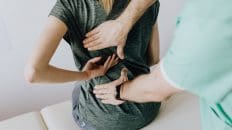
In musculoskeletal medicine, our primary goal is to move beyond symptom management and actively promote healing and restore function. For years, patients with symptomatic knee osteoarthritis (OA) have navigated a treatment paradigm often limited to temporary relief measures. Traditional options like corticosteroid injections, while effective for short-term inflammation control, carry risks with repeated use, including potential cartilage damage. Similarly, viscosupplementation with hyaluronic acid, intended to lubricate the joint, has shown mixed, often modest results. This has left a significant therapeutic gap for a condition that affects millions, progressively degrading their quality of life. As a clinician with dual licensure as a Doctor of Chiropractic (DC) and a Family Nurse Practitioner (FNP-APRN), I integrate evidence-based innovations with a deep understanding of human physiology and biomechanics. I am thrilled to present a comprehensive exploration of one of the most promising advancements in this field: Platelet-Rich Plasma (PRP) therapy.
This educational post provides a deep dive into the science, application, and future of PRP as a leading orthobiologic treatment for knee OA. We will move beyond the surface-level understanding of PRP and delve into the intricate cellular and molecular mechanisms that drive its therapeutic effects. The discussion will begin by contextualizing the immense clinical challenge posed by knee osteoarthritis and examining its pathophysiology, from cartilage degradation to synovial inflammation and subchondral bone changes. We will then pivot to a detailed breakdown of PRP itself—what it is, how it’s prepared, and, most importantly, the powerful cocktail of growth factors and cytokines it contains. I will explain the specific roles of key players like Platelet-Derived Growth Factor (PDGF), Transforming Growth Factor-beta (TGF-?), and Vascular Endothelial Growth Factor (VEGF) in orchestrating a complex healing cascade within the joint.
A central focus of this post will be the robust and growing body of evidence supporting PRP’s efficacy. We will critically analyze landmark studies and meta-analyses comparing PRP head-to-head with corticosteroids and hyaluronic acid, highlighting PRP’s superior long-term outcomes for pain reduction and functional improvement. Crucially, we will explore exciting, emerging research suggesting that PRP may not only alleviate symptoms but also possess chondroprotective and potentially even chondro-regenerative properties. This concept represents a true paradigm shift in OA management. Drawing from my own clinical observations at HealthVoice360.com, I will provide insights into patient selection, treatment protocols, and the real-world impact of this therapy. We will also discuss the importance of proper PRP preparation, the classification of different PRP formulations (e.g., leukocyte-rich vs. leukocyte-poor), and how these variables can influence clinical outcomes. By the end of this comprehensive overview, you will have a thorough understanding of why PRP is no longer considered an experimental novelty but a cornerstone of modern, evidence-based regenerative medicine for knee osteoarthritis.
The Clinical Challenge of Symptomatic Knee Osteoarthritis
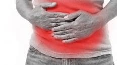
Before we delve into the specifics of regenerative solutions, it is crucial to appreciate the profound and multifaceted nature of symptomatic knee osteoarthritis (OA). As a clinician, I see the impact of this condition daily. It’s not simply “wear and tear” or an inevitable consequence of aging. It is a complex, active disease process involving the entire joint organ—the articular cartilage, the subchondral bone, the synovial membrane, the ligaments, and the menisci.
From a patient’s perspective, the experience begins insidiously. It might start as a mild ache after a long walk, morning stiffness that takes a few minutes to work out, or a subtle “clicking” or “grinding” sensation (crepitus) when bending the knee. Over time, this progresses. The pain becomes more persistent, present even at rest. Swelling (effusion) may occur after activity as the joint becomes inflamed. Simple tasks that were once taken for granted—climbing stairs, getting out of a chair, walking the dog, or kneeling to play with a grandchild—become monumental challenges fraught with pain and instability. This progressive loss of function is what truly defines the burden of symptomatic knee OA. It erodes a person’s independence, limits their social engagement, and can lead to a sedentary lifestyle, which in turn contributes to other comorbidities like obesity, cardiovascular disease, and diabetes.
A Deeper Look at the Pathophysiology of Osteoarthritis
To understand why traditional treatments often fall short and why regenerative approaches like PRP hold so much promise, we must look at what is happening at a biological level within the osteoarthritic joint.
The hallmark of OA is the progressive degradation of articular cartilage. This smooth, resilient tissue covers the ends of the bones in a joint, providing a low-friction surface that allows for effortless movement. It is primarily composed of water, collagen (mainly type II), and proteoglycans (like aggrecan), all of which are produced and maintained by specialized cells called chondrocytes.
In a healthy knee, there is a perfect balance—a state of homeostasis—between the breakdown of old cartilage components and the synthesis of new ones. In osteoarthritis, this balance is disrupted. A cascade of inflammatory and catabolic (breakdown) processes overwhelms the anabolic (build-up) capacity of the chondrocytes.
- Initiating Injury and Inflammation: The process can be triggered by a variety of factors, including acute injury (such as a meniscal tear or ACL rupture), chronic overuse or malalignment, genetic predisposition, or obesity. This initial trigger induces mechanical stress in the chondrocytes.
- Chondrocyte Distress and Catabolic Overdrive: Stressed chondrocytes begin producing pro-inflammatory mediators, such as interleukin-1?. (IL-1?) and tumor necrosis factor-? (TNF-?). These molecules act like alarm bells, but in OA, the alarm never shuts off. They signal the chondrocytes to ramp up production of cartilage-degrading enzymes known as matrix metalloproteinases (MMPs) and aggrecanases (ADAMTS). These enzymes are the “Pac-Men” of the joint, literally chewing through the collagen and proteoglycan framework of the cartilage.
- Synovial Inflammation (Synovitis): The inflammatory mediators and cartilage breakdown products leak into the synovial fluid, irritating the synovial membrane, the thin tissue that lines the joint capsule. This triggers synovitis, an inflammation of the synovium. The inflamed synovium, in turn, produces even more IL-1?, TNF-?, and other inflammatory cytokines, creating a vicious, self-perpetuating cycle of inflammation and degradation. This synovitis is a major source of pain and swelling in the OA knee.
- Cartilage Fibrillation and Erosion: As the matrix breaks down, the cartilage loses its smooth surface. It begins to soften, fray, and develop small cracks—a process called fibrillation. As this continues, the cartilage thins and erodes, eventually exposing the underlying subchondral bone.
- Subchondral Bone Involvement: The bone beneath the cartilage is not a passive bystander. As the cartilage thins, the bone experiences increased stress. It responds by becoming thicker and denser (sclerosis) and by forming bony outgrowths at the joint margins, known as osteophytes or “bone spurs.” These changes can contribute to pain and restricted motion.
This entire process highlights a critical point: knee OA is an inflammatory and catabolic disease. The joint environment is hostile to healing. The very cells responsible for maintaining the cartilage, the chondrocytes, are tricked into destroying it. This is why a treatment that can fundamentally shift this environment from catabolic to anabolic, from pro-inflammatory to anti-inflammatory, is so desperately needed.
The Limitations of Conventional Knee OA Treatments
For decades, our approach to managing this complex disease has been largely palliative. In my clinical practice, I find that while these treatments can be useful, each has significant drawbacks, particularly for long-term management.
Corticosteroid Injections
Intra-articular corticosteroid injections have long been a mainstay for providing rapid, short-term relief from OA pain. Steroids are powerful anti-inflammatory agents. When injected into the knee, they suppress the inflammatory cascade, reducing the production of cytokines such as IL-1? and TNF-?. This calms the synovitis, which in turn reduces pain and swelling. For a patient experiencing an acute flare-up with significant pain and effusion, a steroid injection can be a game-changer, allowing them to sleep through the night or participate in physical therapy.
However, the benefits are notoriously short-lived, typically lasting from a few weeks to, at best, a few months. The underlying degenerative process continues unabated. More concerning is the growing body of evidence regarding the potential long-term harm of repeated steroid injections. Research has shown a correlation between multiple injections and accelerated cartilage loss. The term chondrotoxic is now used to describe this effect. The very treatment used to alleviate symptoms may, in fact, be hastening the progression of the disease it aims to treat. This creates a significant clinical dilemma. As a result, I am very cautious and selective when recommending steroid injections, typically reserving them for specific situations and counseling patients extensively on the risks versus the short-term benefits.
Hyaluronic Acid (HA) Injections (Viscosupplementation)
Viscosupplementation is another common treatment option. The rationale is quite elegant: in an osteoarthritic joint, the synovial fluid loses its viscosity and elasticity. This is because the concentration and molecular weight of naturally occurring hyaluronic acid (HA), a key component of synovial fluid, are reduced. HA acts as a lubricant and a shock absorber for the joint.
HA injections aim to restore these properties by injecting a gel-like HA substance directly into the joint space. The goal is to reduce pain and improve function by lubricating the joint surfaces. While the theory is sound, the clinical results have been inconsistent. Many large-scale, high-quality studies and meta-analyses have shown that HA injections provide, on average, only a small and often not clinically significant benefit over placebo (saline) injections.
While HA is generally considered safe and does not carry the chondrotoxic risk of steroids, its lackluster efficacy for many patients has been disappointing. It doesn’t address the underlying inflammatory and catabolic environment of the joint. In my experience, some patients do report moderate relief, but many find the benefit to be minimal or nonexistent, especially those with more advanced, “bone-on-bone” arthritis. It is a passive, mechanical intervention in a very active, biological disease process. This is precisely where the paradigm shifts toward orthobiologics.
The Orthobiologic Revolution: Introducing Platelet-Rich Plasma (PRP)
This is where my excitement as a clinician truly begins. The field of orthobiologics represents a fundamental shift in our thinking. Instead of introducing synthetic chemicals or lubricants into the joint, we harness the body’s own innate healing capabilities. We use biological substances to create a pro-healing, anti-inflammatory environment that can slow, stop, or even potentially reverse disease-related damage. At the forefront of this revolution for knee OA is Platelet-Rich Plasma (PRP).
I find that PRP is an exceptionally effective and evidence-based modality for my patients with symptomatic knee osteoarthritis. It’s not a new, unproven fad; the science behind it is robust, and the clinical evidence is mounting rapidly.
What Exactly Is Platelet-Rich Plasma?
PRP is, quite simply, a concentration of platelets derived from the patient’s own blood. We all know platelets as the tiny cell fragments that rush to the site of an injury, like a cut, to form a clot and stop bleeding. But their role extends far beyond simple clotting. Platelets are biological powerhouses, acting as miniature storage depots for a vast array of potent signaling molecules, including growth factors and cytokines.
When we are injured, platelets are activated and release these molecules in a concentrated burst at the site of damage. This release initiates and orchestrates the entire healing cascade, recruiting other cells (such as stem and immune cells), promoting the formation of new blood vessels (angiogenesis), and stimulating tissue regeneration.
The core principle of PRP therapy is to capture this natural healing power, concentrate it, and deliver it precisely to a site of chronic injury or degeneration, like an osteoarthritic knee. By doing so, we are essentially telling the joint, “There has been an injury here. It is time to initiate a powerful healing response.”
The PRP Preparation Process: From Blood Draw to Injection
The process is remarkably straightforward and is performed as an outpatient procedure in our clinic.
- Blood Draw: The first step is a simple blood draw, similar to what you would have for routine lab work. We typically draw between 30 and 60 milliliters of blood from a vein in the patient’s arm.
- Centrifugation: The drawn blood is placed in a sterile, specialized kit and then centrifuged. The centrifuge spins the blood at very high speeds, using centrifugal force to separate it into its components based on density.
- The heaviest components, the red blood cells (erythrocytes), settle at the bottom.
- The least dense component, the platelet-poor plasma (PPP), forms the top layer.
- In between these two layers is a thin, yellowish-white layer called the buffy coat. This is the “liquid gold” we are after. The buffy coat contains the highly concentrated platelets and, depending on the system used, a variable number of white blood cells (leukocytes).
- Extraction and Preparation: The platelet-rich plasma, including the buffy coat, is carefully extracted. Depending on the specific protocol and the patient’s condition, we may use a “double spin” technique to achieve an even higher platelet concentration. The final PRP product is a small volume (typically 3-6 mL) of plasma with a platelet concentration 3-8 times that of normal circulating blood.
- Injection: Using sterile technique, the prepared PRP is then injected directly into the knee joint capsule (intra-articularly). In my practice, I often use ultrasound guidance to ensure precise placement of the injection. This allows me to visualize the needle in real time and confirm that the PRP is being delivered directly into the joint space, where it can be most effective, rather than into surrounding soft tissues.
The entire process, from blood draw to injection, typically takes about 30 to 45 minutes. Because the PRP is derived from the patient’s own blood (autologous), the risk of allergic reaction or disease transmission is virtually nonexistent.
The Molecular Power of PRP: Unpacking the Growth Factors
The true magic of PRP lies in the cocktail of bioactive proteins released by the activated platelets. These growth factors are the conductors of the healing orchestra. Let’s examine the key players and their specific roles within the knee joint.
Transforming Growth Factor-beta (TGF-?): The Master Regulator
TGF-? is one of the most important and abundant growth factors in platelets. It’s a “master regulator” with a complex, dual role. It is profoundly influential in tissue regeneration, particularly in cartilage.
- Stimulates Matrix Synthesis: TGF-? directly signals chondrocytes to increase production of the essential building blocks of cartilage: type II collagen and aggrecan. This is the core anabolic effect—it encourages the rebuilding of the cartilage matrix that the OA process has degraded.
- Modulates Inflammation: TGF-? also has potent immunomodulatory effects. It can help suppress the activity of pro-inflammatory cytokines, shifting the joint environment away from chronic inflammation. It plays a delicate balancing act, promoting repair while keeping inflammation in check.
Platelet-Derived Growth Factor (PDGF): The Cell Recruiter and Proliferator
As its name suggests, PDGF is a powerful stimulant for cell growth and division (mitogenesis).
- Cell Proliferation and Migration: PDGF sends out a strong signal that recruits other important cells to the area. It promotes chondrocyte proliferation and attracts mesenchymal stem cells (MSCs) from surrounding tissues, such as the synovium and fat pad. These MSCs are multipotent and can differentiate into new cartilage cells.
- Angiogenesis and Collagen Synthesis: PDGF also promotes angiogenesis and stimulates collagen synthesis, providing the structural framework for new tissue.
Vascular Endothelial Growth Factor (VEGF): The Angiogenesis Factor
VEGF is the primary driver of angiogenesis, the formation of new blood vessels. While articular cartilage itself is avascular (lacks a blood supply), a healthy blood supply to the subchondral bone and surrounding synovial tissues is critical for delivering nutrients and clearing waste products. In the context of healing, a new microvascular network is essential for supporting the metabolic demands of tissue regeneration. VEGF helps ensure that the healing tissues receive the oxygen and nutrients they need to thrive.
Fibroblast Growth Factor (FGF): The Tissue Builder
FGF is another key mitogen that stimulates proliferation in a wide range of cell types, including chondrocytes and fibroblasts. It plays a crucial role in tissue repair and regeneration across the body. In the knee, it contributes to the overall anabolic signal, encouraging cells to build and repair the damaged joint structures.
Insulin-like Growth Factor (IGF-1): The Anabolic Powerhouse
IGF-1 is a potent anabolic growth factor. It works synergistically with other growth factors, such as TGF-? and PDGF, to powerfully stimulate chondrocytes to produce more cartilage matrix. It also helps inhibit programmed cell death (apoptosis) in chondrocytes, thereby preserving the existing cell population.
The Symphony of Healing
It is critically important to understand that these growth factors do not act in isolation. They work in a complex, synergistic symphony. The precise combination and concentration of these factors, released in a specific sequence after PRP injection, create a powerful biological signal that accomplishes several key objectives simultaneously:
- Reduces Inflammation: It downregulates the production of catabolic enzymes (MMPs) and pro-inflammatory cytokines (IL-1?, TNF-?).
- Promotes Anabolism: It stimulates chondrocytes and recruited stem cells to synthesize new cartilage matrix components.
- Alters the Joint Environment: It fundamentally shifts the entire joint milieu from a degenerative, catabolic state to a regenerative, anabolic state.
This multi-pronged mechanism of action is why PRP is so much more effective than single-molecule therapies. A steroid injection only addresses inflammation temporarily. An HA injection only provides temporary lubrication. A PRP injection initiates a cascade of biological events aimed at comprehensive joint repair and homeostasis.
Knee Injury Rehabilitation Story- Video
The Evidence Speaks: PRP vs. Conventional Treatments
As a practitioner committed to evidence-based medicine, I don’t just rely on theory or anecdotal experience. The recommendations I make to my patients are grounded in high-quality scientific research. In the case of PRP for knee OA, the body of evidence has become overwhelmingly positive, especially when compared directly to corticosteroids and hyaluronic acid.
Numerous randomized controlled trials (RCTs) and meta-analyses—the highest levels of medical evidence—have been published in leading orthopedic and sports medicine journals. The consensus from this research is clear and consistent.
Head-to-Head: PRP vs. Corticosteroid Injections
When PRP is compared directly with corticosteroid injections, a distinct pattern emerges.
- Short-Term: In the first few weeks to a month post-injection, both PRP and steroids can provide significant pain relief. In some studies, steroids may even offer slightly faster initial relief due to their potent, rapid anti-inflammatory effects.
- Long-Term: This is where the difference becomes dramatic. The effects of the steroid injection typically wane after 2-3 months, with pain and function levels often returning to baseline. In contrast, the benefits of PRP continue to build over time. Studies consistently show that at 6, 9, and 12 months post-injection, patients in the PRP groups report significantly better outcomes in pain reduction and functional improvement (as measured by standardized scores such as WOMAC, VAS, and IKDC) compared with patients in the steroid groups.
One landmark meta-analysis published in the American Journal of Sports Medicine reviewed data from multiple RCTs. It concluded that intra-articular PRP injections provide greater and longer-lasting clinical improvement for knee OA than corticosteroid injections. The rationale is clear: steroids provide a temporary chemical anti-inflammatory effect, while PRP initiates a long-term biological healing response.
Head-to-Head: PRP vs. Hyaluronic Acid (HA)
The comparison with HA is equally compelling, if not more so, in favor of PRP.
- Multiple high-quality RCTs and meta-analyses have compared single or series of PRP injections against single or series of HA injections.
- The results consistently demonstrate that PRP is statistically and clinically superior to HA in improving pain and function scores at all follow-up points (3, 6, and 12 months).
A 2017 meta-analysis in the Journal of Orthopedic Surgery and Research found that PRP injections yielded better and more durable results than HA injections for knee OA, particularly in patients with less severe (early to moderate) osteoarthritis. This superiority makes sense from a biological standpoint. HA is a passive lubricant, whereas PRP is a biologically active treatment that directly modulates the disease process.
Based on this high-level evidence, I frequently find myself counseling patients who have previously tried steroid or HA injections with limited success. For these individuals, PRP isn’t just another option; it represents a step up to a more advanced, biologically targeted therapy with a much higher probability of providing meaningful, long-term relief.
The Future is Here: Chondroprotective and Regenerative Potential
Perhaps the most exciting and groundbreaking area of PRP research is its potential to do more than manage symptoms. There is emerging literature from both animal models and human clinical studies suggesting that PRP may have a chondroprotective effect—meaning it can slow down or even halt the progression of cartilage degradation.
How Can PRP Protect Cartilage?
The mechanism is tied to its ability to rebalance the joint environment. By suppressing the key inflammatory drivers (IL-1?, TNF-?) and the catabolic enzymes (MMPs) they produce, PRP effectively puts the brakes on the destructive cycle of OA. At the same time, by upregulating the production of matrix components via growth factors such as TGF-? and IGF-1, it strengthens the remaining cartilage, making it more resilient to stress.
Some advanced imaging studies using specialized MRI techniques such as dGEMRIC (delayed gadolinium-enhanced MRI of cartilage) have even hinted at chondro-regenerative potential. These studies have shown improvements in cartilage quality and even small increases in cartilage thickness in patients treated with PRP, which was previously thought impossible in adults. While this research is still in its earlier stages, it is incredibly promising. It suggests that PRP may be one of the first treatments that can truly modify the course of the disease, not just its symptoms.
This is a paradigm shift. For the first time, we are moving from a palliative care model for OA to a genuinely regenerative one. The goal is no longer to make the patient feel better for a few months, but to preserve the joint for years to come, potentially delaying or even preventing the need for major surgery, such as a total knee replacement.
Clinical Insights and Patient Considerations: A Personalized Approach
While the science is compelling, applying it effectively in the clinic requires nuance and a personalized approach. Not every patient with knee pain is an ideal candidate for PRP. At my clinic, the process begins with a thorough evaluation.
Who is a Good Candidate for PRP?
Based on the research and my clinical observations from HealthVoice360.com, the ideal candidates for PRP knee injections typically have:
- Mild to Moderate Osteoarthritis: PRP tends to be most effective in patients with Kellgren-Lawrence Grade I, II, or III OA. In these stages, there is still sufficient cartilage and a resident population of chondrocytes capable of responding to growth factor signaling. The joint environment is diseased but not yet destroyed.
- Failure of Conservative Treatments: Patients who have already tried and failed more conservative measures like physical therapy, NSAIDs, activity modification, and perhaps even steroid or HA injections are often excellent candidates.
- Active Individuals Wishing to Avoid Surgery: I see many patients in their 40s, 50s, and 60s who remain very active but whose knee pain is beginning to limit their lifestyles. They are often deemed “too young” for a knee replacement but are desperate for a solution that will allow them to continue hiking, playing tennis, or simply keeping up with their families. PRP offers a minimally invasive option to bridge this gap.
Conversely, patients with very advanced “bone-on-bone” (Grade IV) arthritis, significant joint deformity, or inflammatory arthropathies such as rheumatoid arthritis may not be the best candidates, as the biological environment may be too severely damaged for PRP to be effective.
The Importance of PRP Formulation: Leukocyte-Rich vs. Leukocyte-Poor
One of the most important and often overlooked aspects of PRP therapy is the composition of the final product. Not all PRP is created equal. A key variable is the concentration of white blood cells (leukocytes).
- Leukocyte-Rich PRP (LR-PRP): This formulation contains a high concentration of both platelets and white blood cells. White blood cells, particularly neutrophils, can release their own set of pro-inflammatory cytokines.
- Leukocyte-Poor PRP (LP-PRP): This formulation is prepared to minimize white blood cells, resulting in a product that is almost entirely platelet-rich.
The debate over which formulation is better is ongoing, but a consensus is beginning to emerge for intra-articular applications like knee OA. The intra-articular space is a unique immune environment. Introducing a high concentration of leukocytes (as in LR-PRP) can sometimes trigger a significant inflammatory flare-up in the days following the injection, causing increased pain and swelling.
For this reason, most leading researchers and clinicians, myself included, now favor Leukocyte-Poor PRP (LP-PRP) for the treatment of knee osteoarthritis. The theory is that LP-PRP provides all the anabolic and regenerative benefits of the platelet-derived growth factors without the potentially counterproductive inflammatory spike from the leukocytes. The goal in OA is to calm the joint, not to provoke a new inflammatory response.
The Post-Injection Protocol: Partnering in the Healing Process
The injection itself is only the beginning of the healing process. What the patient does in the weeks and months following the procedure is critical for maximizing the results. My post-injection protocol is designed to protect the joint while allowing the biological cascade initiated by the PRP to work its magic.
- Initial Rest Period (First 48-72 Hours): I advise patients to take it easy for the first few days. This means avoiding strenuous activity, but gentle movement and walking are encouraged to help circulate the PRP within the joint. Crutches may be used for a day or two if needed for comfort. Critically, I instruct all patients to avoid all non-steroidal anti-inflammatory drugs (NSAIDs) like ibuprofen, naproxen, and celecoxib for at least two weeks before and several weeks after the injection. NSAIDs work by blocking the very inflammatory pathways that PRP aims to modulate for healing. Taking them can counteract the effects of the treatment.
- Gentle Range of Motion (Week 1): Focus on gentle, non-weight-bearing range-of-motion exercises to maintain flexibility and joint health without stressing healing tissues.
- Formal Physical Therapy (Beginning Week 2-3): This is a cornerstone of a successful outcome. The PRP aims to improve the joint’s biological environment, but we must also address the biomechanical issues. A skilled physical therapist will guide the patient through a progressive program of:
-
- Strengthening: Focusing on the quadriceps, hamstrings, glutes, and core muscles to provide dynamic support for the knee joint.
- Neuromuscular Control: Retraining the nerves and muscles to work together efficiently to stabilize the joint during movement.
- Flexibility and Mobility: Restoring normal joint mechanics and addressing any muscle imbalances.
The combination of the biological “boost” from PRP and the biomechanical improvements from physical therapy leads to the best and most durable long-term outcomes. PRP creates a healthier, less painful joint, which in turn allows the patient to participate more effectively in the physical therapy needed to restore true function.
As we move forward, the field of orthobiologics will continue to evolve. We will see more advanced formulations, perhaps combining PRP with other regenerative substances, and our ability to tailor treatments to individual patients will become even more precise. However, as it stands today, the quality of the data and the strength of the evidence are undeniable. For my patients with symptomatic knee osteoarthritis, PRP is no longer a future hope; it is a powerful, effective, and evidence-based modality that is helping them reclaim their lives, one step at a time. It represents a major advancement in our journey toward truly regenerative musculoskeletal care.
Summary, Conclusion, and Key Insights
Summary
This educational post, presented from my perspective as Dr. Alexander Jimenez, DC, APRN, FNP-BC, provided a comprehensive exploration of Platelet-Rich Plasma (PRP) therapy as a modern, evidence-based treatment for symptomatic knee osteoarthritis (OA). We began by defining knee OA not merely as “wear and tear,” but as a complex, active disease process involving cartilage degradation, chronic synovial inflammation (synovitis), and subchondral bone changes, all driven by a self-perpetuating cycle of catabolic and pro-inflammatory mediators. We contrasted this with the limitations of conventional treatments: corticosteroid injections offer only temporary relief and carry a risk of chondrotoxicity. At the same time, hyaluronic acid (viscosupplementation) has demonstrated inconsistent, often minimal efficacy.
The discussion focused on PRP as a leading orthobiologic solution. We detailed the autologous preparation process, from a simple blood draw to centrifugation and extraction of the platelet-rich buffy coat. The primary mechanism of action was explained as the “symphony of healing” orchestrated by powerful growth factors released from activated platelets. We examined the specific roles of key molecules such as Transforming Growth Factor-beta (TGF-?) in matrix synthesis, Platelet-Derived Growth Factor (PDGF) in cell recruitment, and Insulin-like Growth Factor (IGF-1) for its potent anabolic effects. This biological cascade shifts the joint environment from a degenerative, catabolic state to a regenerative, anabolic one. We critically analyzed high-level evidence from meta-analyses and randomized controlled trials, which consistently show PRP to be superior to both corticosteroids and hyaluronic acid for long-term pain relief and functional improvement at 6-, 9-, and 12-month follow-ups. Furthermore, we explored exciting, emerging research suggesting PRP may be chondroprotective, potentially slowing disease progression. Finally, we covered crucial clinical considerations, including patient selection (favoring mild-to-moderate OA), the importance of using Leukocyte-Poor PRP (LP-PRP) for intra-articular injections to minimize inflammatory reactions, and the essential role of a structured post-injection protocol centered on physical therapy.
Conclusion
The accumulated scientific evidence and extensive clinical experience have firmly established Platelet-Rich Plasma therapy as a cornerstone in the non-operative management of symptomatic knee osteoarthritis. It represents a significant evolution beyond palliative treatments that only mask symptoms. By harnessing the body’s own healing potential, PRP directly targets the underlying pathophysiology of OA, reducing inflammation, stimulating anabolic processes, and fundamentally altering the joint milieu in favor of repair and homeostasis. Head-to-head comparisons with traditional injectables such as corticosteroids and hyaluronic acid are no longer in question; the data clearly demonstrate PRP’s superior, more durable outcomes. The potential for genuine disease modification and chondroprotection moves PRP from the realm of symptom management into the exciting new territory of regenerative medicine. For clinicians and patients alike, PRP offers a safe, effective, and evidence-based strategy to improve function, reduce pain, and enhance quality of life, potentially delaying or even obviating the need for major joint replacement surgery.
Key Insights
- PRP is Biologically Active, Not Passive: Unlike hyaluronic acid (a lubricant) or corticosteroids (chemical anti-inflammatories), PRP is a biological therapy that initiates a complex healing cascade within the joint, promoting a long-term shift toward an anabolic, regenerative state.
- Evidence Demonstrates Superior Long-Term Efficacy: High-level evidence from multiple meta-analyses confirms that PRP provides significantly better, longer-lasting pain relief and functional improvement than corticosteroid and hyaluronic acid injections at 6- and 12-month follow-ups.
- Formulation Matters: Leukocyte-Poor is Preferred. For intra-articular treatment of osteoarthritis, Leukocyte-Poor PRP (LP-PRP) is generally favored over Leukocyte-Rich (LR-PRP) to avoid a post-injection inflammatory flare and to focus on the anabolic effects of growth factors.
- PRP May Be Disease-Modifying: Emerging research suggests PRP has a chondroprotective effect, meaning it can slow the progression of cartilage breakdown. This represents a paradigm shift from simple symptom relief to true modification of the disease course.
- Treatment is a Partnership: The success of PRP therapy is maximized when paired with a dedicated post-procedure rehabilitation program. Physical therapy to address strength, stability, and biomechanics is a critical component for achieving optimal, lasting results.
References
- Laudy, A. B., Bakker, E. W., Rekers, M., & Moen, M. H. (2015). Efficacy of platelet-rich plasma injections in osteoarthritis of the knee: a systematic review and meta-analysis. British Journal of Sports Medicine, 49(10), 657-665.
- Meheux, C. J., McCulloch, P. C., Lintner, D. M., Varner, K. E., & Harris, J. D. (2016). Efficacy of intra-articular platelet-rich plasma injections in knee osteoarthritis: a systematic review. Arthroscopy: The Journal of Arthroscopic & Related Surgery, 32(3), 495-505.
- Shen, L., Yuan, T., Chen, S., Xie, X., & Zhang, C. (2017). The temporal effect of platelet-rich plasma on pain and physical function in the treatment of knee osteoarthritis: systematic review and meta-analysis of randomized controlled trials. Journal of Orthopedic Surgery and Research, 12(1), 16.
- Dai, W. L., Zhou, A. G., Zhang, H., & Zhang, J. (2017). Efficacy of platelet-rich plasma in the treatment of knee osteoarthritis: a meta-analysis of randomized controlled trials. Arthroscopy: The Journal of Arthroscopic & Related Surgery, 33(3), 659-670.e1.
- Filardo, G., Kon, E., Buda, R., Timoncini, A., Di Martino, A., Cenacchi, A., … & Marcacci, M. (2011). Platelet-rich plasma intra-articular knee injections for the treatment of degenerative cartilage lesions and osteoarthritis. Knee Surgery, Sports Traumatology, Arthroscopy, 19(4), 528-535.
- Sánchez, M., Guadilla, J., Fiz, N., & Andia, I. (2012). Ultrasound-guided platelet-rich plasma injections for the treatment of osteoarthritis of the knee. Rheumatology, 51(1), 144-150.
- Riboh, J. C., Saltzman, B. M., Yanke, A. B., & Cole, B. J. (2016). Effect of leukocyte concentration on the efficacy of platelet-rich plasma in the treatment of knee osteoarthritis. The American Journal of Sports Medicine, 44(3), 792-800.
Keywords
Knee Osteoarthritis, Platelet-Rich Plasma, PRP, Orthobiologics, Regenerative Medicine, Knee Pain, Growth Factors, Chondroprotection, Corticosteroid Injections, Hyaluronic Acid, Viscosupplementation, Non-operative treatment, Dr. Alexander Jimenez, Evidence-Based Medicine, Leukocyte-Poor PRP, Physical Therapy, Joint Preservation.
Disclaimer: The information contained in this post is for educational and informational purposes only and is not intended as health or medical advice. It is not a substitute for professional medical advice, diagnosis, or treatment. Always seek the advice of your physician or other qualified health provider with any questions you may have regarding a medical condition or treatment and before undertaking a new health care regimen. Never disregard professional medical advice or delay in seeking it because of something you have read on this website.
Patient-Specific Advice Disclaimer: All individuals are unique, and the appropriate medical treatment for one person may not be suitable for another. The content presented here reflects general principles and findings in the medical field. Everyone should obtain personalized recommendations for their specific health situation directly from their qualified medical providers, who can assess their complete medical history and current condition.
General Disclaimer
Professional Scope of Practice *
The information herein on "Knee Osteoarthritis Overview and PRP Therapy Insights" is not intended to replace a one-on-one relationship with a qualified health care professional or licensed physician and is not medical advice. We encourage you to make healthcare decisions based on your research and partnership with a qualified healthcare professional.
Blog Information & Scope Discussions
Welcome to El Paso's Premier Wellness and Injury Care Clinic & Wellness Blog, where Dr. Alex Jimenez, DC, FNP-C, a Multi-State board-certified Family Practice Nurse Practitioner (FNP-BC) and Chiropractor (DC), presents insights on how our multidisciplinary team is dedicated to holistic healing and personalized care. Our practice aligns with evidence-based treatment protocols inspired by integrative medicine principles, similar to those found on this site and our family practice-based chiromed.com site, focusing on restoring health naturally for patients of all ages.
Our areas of multidisciplinary practice include Wellness & Nutrition, Chronic Pain, Personal Injury, Auto Accident Care, Work Injuries, Back Injury, Low Back Pain, Neck Pain, Migraine Headaches, Sports Injuries, Severe Sciatica, Scoliosis, Complex Herniated Discs, Fibromyalgia, Chronic Pain, Complex Injuries, Stress Management, Functional Medicine Treatments, and in-scope care protocols.
Our information scope is multidisciplinary, focusing on musculoskeletal and physical medicine, wellness, contributing etiological viscerosomatic disturbances within clinical presentations, associated somato-visceral reflex clinical dynamics, subluxation complexes, sensitive health issues, and functional medicine articles, topics, and discussions.
We provide and present clinical collaboration with specialists from various disciplines. Each specialist is governed by their professional scope of practice and their jurisdiction of licensure. We use functional health & wellness protocols to treat and support care for musculoskeletal injuries or disorders.
Our videos, posts, topics, and insights address clinical matters and issues that are directly or indirectly related to our clinical scope of practice.
Our office has made a reasonable effort to provide supportive citations and has identified relevant research studies that support our posts. We provide copies of supporting research studies upon request to regulatory boards and the public.
We understand that we cover matters that require an additional explanation of how they may assist in a particular care plan or treatment protocol; therefore, to discuss the subject matter above further, please feel free to ask Dr. Alex Jimenez, DC, APRN, FNP-BC, or contact us at 915-850-0900.
We are here to help you and your family.
Blessings
Dr. Alex Jimenez DC, MSACP, APRN, FNP-BC*, CCST, IFMCP, CFMP, ATN
email: coach@elpasofunctionalmedicine.com
Multidisciplinary Licensing & Board Certifications:
Licensed as a Doctor of Chiropractic (DC) in Texas & New Mexico*
Texas DC License #: TX5807, Verified: TX5807
New Mexico DC License #: NM-DC2182, Verified: NM-DC2182
Multi-State Advanced Practice Registered Nurse (APRN*) in Texas & Multistate
Multistate Compact RN License by Endorsement (42 States)
Texas APRN License #: 1191402, Verified: 1191402 *
Florida APRN License #: 11043890, Verified: APRN11043890 *
* Prescriptive Authority Authorized
ANCC FNP-BC: Board Certified Nurse Practitioner*
Compact Status: Multi-State License: Authorized to Practice in 40 States*
Graduate with Honors: ICHS: MSN-FNP (Family Nurse Practitioner Program)
Degree Granted. Master's in Family Practice MSN Diploma (Cum Laude)
Dr. Alex Jimenez, DC, APRN, FNP-BC*, CFMP, IFMCP, ATN, CCST
My Digital Business Card
RN: Registered Nurse
APRNP: Advanced Practice Registered Nurse
FNP: Family Practice Specialization
DC: Doctor of Chiropractic
CFMP: Certified Functional Medicine Provider
MSN-FNP: Master of Science in Family Practice Medicine
MSACP: Master of Science in Advanced Clinical Practice
IFMCP: Institute of Functional Medicine
CCST: Certified Chiropractic Spinal Trauma
ATN: Advanced Translational Neutrogenomics